 血管に関するMeSH用語の体系的学習
Last Update 2009/05/20| 血管の解剖学用語 | |||
(1) 動脈と静脈 (2) 心臓の4つの部屋につく血管 (3)動脈(Arteries)のMeSH用語一覧 (4)大動脈弓の枝(1):腕頭動脈を探る (5)腕頭動脈(Brachiocephalic Trunk)からの流れ(概略) (6)大動脈弓の枝[2]:総頸動脈を探る:外頸動脈と内頸動脈 (7)顎動脈と眼動脈 (8)映画『ミクロの決死圏』:総頸動脈:注入部位の取り違え? (9)長嶋茂雄氏の梗塞部位:中大脳動脈 (10)大動脈弓の枝[3]鎖骨下動脈を探る:[1]椎骨動脈(ついこつどうみゃく) (11)大動脈弓の枝[3]鎖骨下動脈を探る:[2]脳底動脈(のうていどうみゃく) (12)大脳動脈(Cerebral Arteries)の構成 (13)鎖骨下動脈につづく動脈[1]腋窩動脈(Axillary Artery)と上腕動脈(Brachial Artery) (14)鎖骨下動脈につづく動脈[2]橈骨動脈(Radial Artery)と尺骨動脈(Ulnar Artery) (15)大動脈の構成 (16)腹腔動脈 (17)脾動脈 (18)腎動脈 (19)総肝動脈と固有肝動脈 (20)腸間膜動脈:上腸間膜動脈と下腸間膜動脈[1] (21)腸間膜動脈:上腸間膜動脈と下腸間膜動脈[2] (22)腸骨動脈 (23)大腿動脈(Femoral Artery)と膝窩動脈(Popliteal Artery) (24)脛骨動脈(けいこつどうみゃく) (25)臍動脈(さいどうみゃく) (26)腹壁動脈(ふくへきどうみゃく) (27)静脈弁 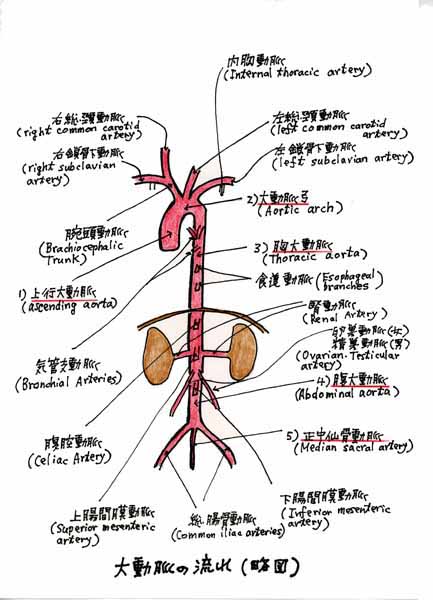 (1) 動脈と静脈 ●動脈(Arteries)とは、血液を心臓から、からだの各部に遠心的(centrifugal)に送り出す血管です。 ●これに対して、静脈(Veins)とは、血液をからだの各部から心臓に向って求心的(centripetal)に 送り返す血管のことをいいます。 ●右心室(right ventricule)からは、肺に向かう肺動脈[幹](pulmonary trunk)が出ていますが、これ には静脈血が流れています。 ●左心房(left atrium)には、肺で動脈血となった血液を受け取る肺静脈(pulmonary veins)が接続 しています。 ●また、胎生期の臍動脈(さいどうみゃく)(umbilical arteries)には静脈血と動脈血の混合血が流れ、 臍静脈(umbilical vein)には動脈血が流れています。 ●Blood Vessels(血管) (http://www.nlm.nih.gov/cgi/mesh/2004/MB_cgi?term=Blood+Vessels&field=entry) (http://www.ncbi.nlm.nih.gov:80/entrez/query.fcgi?cmd=Retrieve&db=mesh&list_uids=68001808&dopt=Full) Scope Note : Any of the tubular vessels conveying the blood (arteries, arterioles, capillaries, venules, and veins). 血管とは、血液を運ぶ管状脈管をいう。 1.Arteries(動脈) (http://www.nlm.nih.gov/cgi/mesh/2004/MB_cgi?term=Arteries&field=entry) (http://www.ncbi.nlm.nih.gov:80/entrez/query.fcgi?cmd=Retrieve&db=mesh&list_uids=68001158&dopt=Full) Scope Note : The vessels carrying blood away from the heart. 動脈とは、心臓から血液を運び出す血管をいう。 2.Veins(静脈) (http://www.nlm.nih.gov/cgi/mesh/2004/MB_cgi?term=Veins&field=entry) (http://www.ncbi.nlm.nih.gov:80/entrez/query.fcgi?cmd=Retrieve&db=mesh&list_uids=68014680&dopt=Full) Scope Note : The vessels carrying blood toward the heart. 静脈とは、心臓へ血液を運びもどす血管をいう。 ・・・・・・・・・・・・・・・・・・・・・・・・・・・ (参 考) ←出る: ←入る: ←←←←←←←←←←←←←← 心 臓 ←←←←←←←←←←←←← 動 脈(Arteries) 静 脈(Veins) ========================================================== 部屋:右心室(肺動脈)(静脈血) 部屋:右心房(大静脈)(静脈血) --------------------------------------------------------------------------------------------------- 部屋:左心室(大動脈)(動脈血) 部屋:左心房(肺静脈)(動脈血) --------------------------------------------------------------------------------------------------- ・・・・・・・・・・・・・・・・・・・・・・・・・・・ (2) 心臓の4つの部屋につく血管 [右心系] 1. 右心房 a.上大静脈(Vena Cava, Superior) (http://www.nlm.nih.gov/cgi/mesh/2004/MB_cgi?term=Vena+Cava,+Superior&field=entry) (http://www.ncbi.nlm.nih.gov:80/entrez/query.fcgi?cmd=Retrieve&db=mesh&list_uids=68014683&dopt=Full) Scope Note The venous trunk which returns blood from the head, neck, upper extremities and chest. 上大静脈とは、頭部、頚部、上肢および胸部からの血液をもどす静脈[幹]をいう。 b.下大静脈(Vena Cava, Inferior) (http://www.nlm.nih.gov/cgi/mesh/2004/MB_cgi?term=Vena+Cava,+Inferior&field=entry) (http://www.ncbi.nlm.nih.gov:80/entrez/query.fcgi?cmd=Retrieve&db=mesh&list_uids=68014682&dopt=Full) Scope Note The venous trunk which receives blood from the lower extremities and from the pelvic and abdominal organs. 下大静脈とは、下肢、骨盤および腹腔器官からの血液を受け取る静脈[幹]をいう。 c.冠状静脈洞(coronary sinus) ●冠状静脈洞は、右心房の後壁の下大静脈の内側にあり、心臓壁の血液を集めます。 ●MeSHでは、coronary sinusは、MeSH用語(Main Headings)ではありません。また、エントリー・タームにも登録されていません。 2. 右心室 肺動脈[幹](Pulmonary Artery) (http://www.nlm.nih.gov/cgi/mesh/2004/MB_cgi?term=Pulmonary+Artery&field=entry) (http://www.ncbi.nlm.nih.gov:80/entrez/query.fcgi?cmd=Retrieve&db=mesh&list_uids=68011651&dopt=Full) Scope Note The short wide vessel arising from the conus arteriosus of the right ventricle and conveying unaerated blood to the lungs. 肺動脈[幹]とは、右心室の動脈円錐(conus arteriosus)から起き、肺へ酸素化されていない血液を運ぶ、短く幅広い血管をいう。 ●肺動脈[幹]の長さは、4〜5cmで、第4胸椎の高さで、右肺動脈(right pulmonary artery)と左肺動脈(left pulmonary artery)とに分かれます。 ↑ ・・・・・・・・・・・・・・・・・・・・・・・・・・・・・・・・・・・・・・・・・・・・・・・・・・・・・・・・・・・・・・・・・・・・ ↓ [左心系] 3. 左心房 肺静脈(pulmonary veins) (http://www.nlm.nih.gov/cgi/mesh/2004/MB_cgi?term=Pulmonary+Veins&field=entry) (http://www.ncbi.nlm.nih.gov:80/entrez/query.fcgi?cmd=Retrieve&db=mesh&list_uids=68011667&dopt=Full) Scope Note The veins that return the oxygenated blood from the lungs to the left atrium of the heart. 肺静脈とは、肺から左心房へ酸素化されて血液をもどす静脈をいう。 4. 左心室 大動脈(aorta) (http://www.nlm.nih.gov/cgi/mesh/2004/MB_cgi?term=Aorta&field=entry) (http://www.ncbi.nlm.nih.gov:80/entrez/query.fcgi?cmd=Retrieve&db=mesh&list_uids=68001011&dopt=Full) Scope Note The main trunk of the systemic arteries. 大動脈とは、体循環・動脈の本幹をいう。 [Coffee Break Test] 右心房、右心室、左心房、左心室 それぞれの英語の解剖学名とMeSH用語ではなんというかを確認しておきましょう。 (3)動脈(Arteries)のMeSH用語一覧 ●MeSH用語のArteriesのカテゴリーは、基本的にABC順に配列されていて、大動脈(Aorta)の枝によって、 解剖学的に階層を分けているという訳ではありません。 (http://www.nlm.nih.gov/cgi/mesh/2004/MB_cgi?term=Arteries&field=entry) ●Aorta(大動脈)、Carotid Arteries(頸動脈)、Cerebral Arteries(大脳動脈)、Mesenteric Arteries(腸間膜動脈)、 Thoracic Arteries(胸動脈)の5つのMeSH用語だけは、下位の階層を展開しています。 (http://www.ncbi.nlm.nih.gov:80/entrez/query.fcgi?cmd=Retrieve&db=mesh&list_uids=68001158&dopt=Full) ●体循環の本幹である大動脈(Aorta)は、上行大動脈(ascending aorta)、大動脈弓(aortic arch)、 胸大動脈(Aorta, Thoracic)、腹大動脈(Aorta, Abdominal)と進み正中仙骨動脈(median sacral artery) に終ります。 ●腹大動脈(Aorta, Abdominal)は、第4腰椎で右総腸骨動脈(right common Iliac Artery)と 左総腸骨動脈(left common Iliac Artery)に分枝します。 ●左心室からはじまる大動脈[幹](Aorta)は、その途中で、樹木の枝のように血管の枝を出しています。 ● 1.上行大動脈(ascending aorta)からは冠状動脈(right and left coronary arteries)、 2.大動脈弓(aortic arch)からは腕頭動脈(Brachiocephalic Trunk)・ 左総頸動脈(left common carotid artery)・左鎖骨下動脈(left subclavian artery)、 3.胸大動脈(thoracic aorta)からは気管支動脈(Bronchial Arteries)、 4.腹大動脈(Aorta, Abdominal)からは腹腔動脈(Celiac Artery)・腸間膜動脈(Mesenteric Arteries)・ 腎動脈(Renal Artery)・腰動脈(lumbar artery)の枝を出しています。 ●MeSH用語のArteriesを、大動脈(Aorta)の枝の流れのなかで、分類し直してみると、 さらに理解が深まるのではないでしょうか。すこしづつ動脈のなかを旅してみたいと思います。 Arteries (動脈) Aorta(大動脈)(だいどうみゃく) Arterioles (細動脈)(さいどうみゃく) Axillary Artery (腋窩動脈)(えきかどうみゃく) Basilar Artery (脳底動脈)(のうていどうみゃく) Brachial Artery (上腕動脈)(じょうわんどうみゃく) Brachiocephalic Trunk(腕頭動脈)(わんとうどうみゃく) Bronchial Arteries(気管支動脈)(きかんしどうみゃく) Carotid Arteries (頸動脈)(けいどうみゃく) Celiac Artery(腹腔動脈)(ふくくうどみゃく) Cerebral Arteries (大脳動脈)(だいのうどうみゃく) Ciliary Arteries (毛様体動脈)(もうようたいどうみゃく) Coronary Vessels(冠状血管)(かんじょうけっかん) Epigastric Arteries (腹壁動脈)(ふくへきどうみゃく) Femoral Artery (大腿動脈)(だいたいどうみゃく) Gastroepiploic Artery (胃大網動脈)(いだいもうどうみゃく) Hepatic Artery (肝動脈)(かんどうみゃく) Iliac Artery (腸骨動脈)(ちょうこつどうみゃく) Maxillary Artery (顎動脈)(がくどうみゃく) Meningeal Arteries (硬膜動脈)(こうまくどうみゃく) Mesenteric Arteries(腸間膜動脈)(ちょうかんまくどうみゃく) Ophthalmic Artery (眼動脈)(がんどうみゃく) Popliteal Artery (膝窩動脈)(しつかどうみゃく) Pulmonary Artery (肺動脈)(はいどうみゃく) Radial Artery (橈骨動脈)(とうこつどうみゃく) Renal Artery (腎動脈)(じんどうみゃく) Retinal Artery (網膜動脈)(もうまくどうみゃく) Splenic Artery (脾動脈)(ひどうみゃく) Subclavian Artery(鎖骨下動脈)(さこつかどうみゃく) Thoracic Arteries (胸動脈)(きょうどうみゃく) Tibial Arteries (脛骨動脈)(けいこつどうみゃく) Ulnar Artery (尺骨動脈)(しゃくこつどうみゃく) Umbilical Arteries (臍動脈)(さいどうみゃく) Vertebral Artery (椎骨動脈)(ついこつどうみゃく) (注) 茶色の動脈は、大動脈(Aorta)から直接出るか分枝する動脈を示します。 (4)大動脈弓の枝(1):腕頭動脈を探る ●腕頭動脈(Brachiocephalic Trunk)は、大動脈弓から出る最も大きな動脈幹で、長さは4〜5cmあり、右総頸動脈と右鎖骨下動脈に分かれます。 (http://www.nlm.nih.gov/cgi/mesh/2004/MB_cgi?term=Brachiocephalic+Trunk&field=entry) (http://www.ncbi.nlm.nih.gov:80/entrez/query.fcgi?cmd=Retrieve&db=mesh&list_uids=68016122&dopt=Full) Scope Note : The first and largest artery branching from the aortic arch. It distributes blood to the right side of the head and neck and to the right arm 腕頭動脈は、大動脈弓から出る最初の最も大きな動脈である。頭部と頚部の右側と右腕に血液を導く。 Previous Indexing : Innominate Artery (1966-1990) 以前は、無名動脈(1966-1990)の名称で索引された。 ●右総頸動脈は、内頸動脈と外頸動脈とに分かれ、右鎖骨下動脈は椎骨動脈、内胸動脈、甲状頸動脈、肋頸動脈とに分かれます。 I.右総頸動脈(right common carotid artery)(Carotid Artery, Common) a.内頸動脈(internal carotid artery)(Carotid Artery, Internal) 眼動脈(ophthalmic artery)(Ophthalmic Artery) 毛様体動脈(ciliary artery)(Ciliary Arteries) 大脳動脈(cerebral arteries)(Cerebral Arteries) b.外頸動脈(external carotic artery)(Carotid Artery, External) 顎動脈(maxillary artery)(Maxillary Artery) II.右鎖骨下動脈(right subclavian artery)(Subclavian Artery) a.椎骨動脈(vertebral artery)(Vertebral Artery) 脳底動脈(basilar artery)(Basilar Artery) b.内胸動脈(internal thoracic artery)(Thoracic Arteries) c.甲状頸動脈(thyrocervical artery) d.肋頸動脈(costocervical trunk) (注) 茶色の英語名はMeSH用語を示します。 (Coffee Break Test) Meningeal Artery(硬膜動脈)・Retinal Artery(網膜動脈)は、どの位置に入るか考えてみましょう。 (5)腕頭動脈(Brachiocephalic Trunk)からの流れ(概略) (http://www.nlm.nih.gov/cgi/mesh/2004/MB_cgi?term=Brachiocephalic+Trunk&field=entry) 脳底 大脳 眼→毛様体 ↑ ↑ ↑ ↑ 顎 ↑ ↑ ↑ ↑ ―――― ↑ ↑ ↑ ↑ 外頸 内頸 ↑ ↑ ↑ ↑ ――――――― ↑ ↑ ↑ ↑ 甲状頸 椎骨 肋頸 ↑ ↑ ↑ ↑ ↑ ↑ ↑ ↑ ↑ ――――――――― ↑ ↑ ↓ ↓ ↑ ↑ 内胸 ↑ ↑ ↑ ↑ ↑ ↑ ↑ 右鎖骨下 右総頸 ↑ ↑ ↑ ↑ ↑ ↑ ―――――――――――――――――― ↑ ↑ ↑ 腕頭 左総頸 左鎖骨下 ↑ ↑ ↑ ――――――――――――――――――――― ↑ ↑ ↑ 大動脈弓 (Coffee Break Test) ・日本語名となっている動脈名を英語名とMeSH用語で置き換えてみましょう。 ・硬膜動脈・網膜動脈を流れ図に書き込んで、流れ図を訂正してみましょう。 (6)大動脈の枝[2]:総頸動脈を探る:外頸動脈と内頸動脈 ●総頸動脈(Carotid Artery, Common)は、頸と頭とに分布する動脈で、左総頸動脈は、直接、大動脈弓から起り、 右総頸動脈は腕頭動脈(Brachiocephalic Trunk)から起ります。 ●総頸動脈は上行して甲状軟骨の上縁の高さで、外頸動脈(Carotid Artery, External)と内頸動脈(Carotid Artery, Internal)とに分かれます。 ●外頸動脈は、上甲状腺動脈(superior thyroid artery)、上行咽頭動脈(ascending pharyngeal artery)、 舌動脈(lingual artery)、顔面動脈(facial artery)、後頭動脈(occipital artery)、後耳介動脈(posterior auricular artery)、浅側頭動脈(superficial temporal artery)(終枝)、顎動脈(Maxillary Artery)(終枝)に 分かれます。顎動脈からは、中硬膜動脈(middle Meningeal artery)(Meningeal Arteries)が出ます。 ●内頸動脈の起始部は、ややふくらんでいて、ここを頸動脈洞(Carotid Sinus)といい、圧迫(刺激)することに よって徐脈と降圧が起こる頸動脈症状が知られています。 ●内頸動脈は、主として脳・眼窩・前頭部に広がり、眼動脈(Ophthalmic Artery>[網膜中心動脈(central Retinal Artery)・毛様体動脈(Ciliary Arteries)]、前大脳動脈(anterior cerebral artery)(Cerebral Arteries)、 中大脳動脈(middle cerebral artery)(Cerebral Arteries)(内頸動脈の終枝)、後交通動脈(posterior communicating artery)に分かれます。 ●MeSHでは、頸動脈(Carotid Arteries)の下位に総頸動脈(Carotid Artery, Common)と頸動脈洞 (Carotid Sinus)とが位置付けられています。 Carotid Arteries(頸動脈) Carotid Artery, Common(総頸動脈) Carotid Artery, Externa(外頸動脈) Carotid Artery, Internal(内頸動脈) Carotid Sinus (頸動脈洞) 1.Carotid Artery, Common(総頸動脈) (http://www.nlm.nih.gov/cgi/mesh/2004/MB_cgi?term=Carotid+Artery,+Common&field=entry) (http://www.ncbi.nlm.nih.gov:80/entrez/query.fcgi?cmd=Retrieve&db=mesh&list_uids=68017536&dopt=Full) Scope Note : The two principal arteries supplying the structures of the head and neck. They ascend in the neck, one on each side, and at the level of the upper border of the thyroid cartilage, each divides into two branches, the external (CAROTID ARTERY, EXTERNAL) and internal (CAROTID ARTERY, INTERNAL) carotid arteries. 総頸動脈は、頭と頸の部位に血液を供給する2本の重要な動脈であり、それらは、左右1本づつ頸の なかを上行して、甲状軟骨の上縁の部分で、それぞれに2本の枝、すなわち外頸動脈(CAROTID ARTERY, EXTERNAL)と内頸動脈(CAROTID ARTERY, INTERNAL)とに分かれる。 1-1. Carotid Artery, External(外頸動脈) (http://www.nlm.nih.gov/cgi/mesh/2004/MB_cgi?term=Carotid+Artery,+External&field=entry) (http://www.ncbi.nlm.nih.gov:80/entrez/query.fcgi?cmd=Retrieve&db=mesh&list_uids=68002342&dopt=Full) Scope Note : Branch of the common carotid artery which supplies the exterior of the head, the face, and the greater part of the neck. 外頸動脈は、総頸動脈(Carotid Artery, Common)の枝で、頭の外面、顔、そして頸の大部分に血液を供給する。 1-2.Carotid Artery, Internal(内頸動脈) (http://www.nlm.nih.gov/cgi/mesh/2004/MB_cgi?term=Carotid+Artery,+Internal&field=entry) (http://www.ncbi.nlm.nih.gov:80/entrez/query.fcgi?cmd=Retrieve&db=mesh&list_uids=68002343&dopt=Full) Scope Note : Branch of the common carotid artery which supplies the anterior part of the brain, the eye and its appendages, the forehead and nose. 内頸動脈は、総頸動脈(Carotid Artery, Common)の枝で、前頭部、眼とその付属器官および額と鼻に血液を供給する。 2.Carotid Sinus(頸動脈洞) (http://www.nlm.nih.gov/cgi/mesh/2004/MB_cgi?term=Carotid+Sinus&field=entry) (http://www.ncbi.nlm.nih.gov:80/entrez/query.fcgi?cmd=Retrieve&db=mesh&list_uids=68002346&dopt=Full) Scope Note : The dilated portion of the common carotid artery at its bifurcation into external and internal carotids. It contains baroreceptors which, when stimulated, cause slowing of the heart, vasodilatation, and a fall in blood pressure. 頸動脈洞は、外頸動脈と内頸動脈との分岐部にある総頸動脈の膨隆をいう。そこには、圧受容器があって、 刺激によって、除脈、血管拡張、降圧が起こる。 ●スコープ・ノートでは、頸動脈洞(Carotid Sinus)を総頸動脈(Carotid Artery, Common)の分岐部の膨隆として、 内頸動脈(Carotid Artery, Internal)の起始部の膨隆とはしていません。 ●そこで、MeSHの参考図書である『グレイ解剖学』でCarotid Sinusをどのように定義しているかをみてみることに しました。 頸動脈洞(Carotid Sinus):「総頸動脈の末端から内頸動脈起始部にかけてのややふくらんだ部、 あるいは内頸動脈そのものに限局した1cm程のふくらみである。」(『グレイ解剖学』) (7)顎動脈と眼動脈 ●顎動脈は外頸動脈の枝で、眼動脈は内頸動脈の枝です。 外頸動脈 →→→→→→ 顎動脈 →→→→→ 中硬膜動脈 内頸動脈 →→→ 眼動脈 →→→ 網膜中心動脈 ↓ ↓ →→ 毛様体動脈 (Coffee break test) 日本語となっている動脈名の英語名とMeSH用語を考えてみましょう。 1)顎動脈(Maxillary Artery) ○「顎動脈は、外頸動脈の終枝の1つで、主として顔面の深部に広がる。下顎頸の後下側で浅側頭動脈: から分かれると、強く迂回して下顎枝の内側に沿って、ほとんど水平に前進し、翼口蓋窩まで進む」 (『日本人体解剖学』金子丑之助原著 南山堂) Maxillary Artery(顎動脈) (http://www.nlm.nih.gov/cgi/mesh/2004/MB_cgi?term=Maxillary+Artery&field=entry) (http://www.ncbi.nlm.nih.gov:80/entrez/query.fcgi?cmd=Retrieve&db=mesh&list_uids=68008438&dopt=Full) Scope Note : A branch of the external carotid artery which distributes to the deep structures of the face (internal maxillary) and to the side of the face and nose (external maxillary). 顎動脈は外頸動脈の枝で顔面の深部に広がる動脈(内顎)(注)と顔面および鼻腔壁に広がる動脈(外顎)とがある。 ●(注)『グレイ解剖学』にinternal maxillary artery(内顎動脈)という用語の記載があります。 これは、顎動脈(maxillary artery)の同義語として使われています。 2) 眼動脈(Ophthalmic Artery) ●眼動脈には、網膜動脈系と毛様体動脈系とがあります。 「視神経とともに視神経管を通って眼窩にはいり、多数の枝に分かれて眼窩の内容(眼球・涙腺・結膜)・ 眼瞼・前頭部・鼻腔壁などに分布する」(『人体解剖学』藤田恒太郎著 南江堂) Ophthalmic Artery(眼動脈) (http://www.nlm.nih.gov/cgi/mesh/2004/MB_cgi?term=Ophthalmic+Artery&field=entry) (http://www.ncbi.nlm.nih.gov:80/entrez/query.fcgi?cmd=Retrieve&db=mesh&list_uids=68009880&dopt=Full) Scope Note : Artery originating from the internal carotid artery and distributing to the eye, orbit and adjacent facial structures. 眼動脈は、内頸動脈にはじまる動脈で、眼、眼窩および隣接の顔面部位に広がる。 2.1 Retinal Artery(網膜動脈) (http://www.nlm.nih.gov/cgi/mesh/2004/MB_cgi?term=Retinal+Artery&field=entry) (http://www.ncbi.nlm.nih.gov:80/entrez/query.fcgi?cmd=Retrieve&db=mesh&list_uids=68012161&dopt=Full) Scope Note : Central retinal artery and its branches. It arises from the ophthalmic artery, pierces the optic nerve and runs through its center, enters the eye through the porus opticus and branches to supply the retina. 網膜動脈は、網膜中心動脈とその枝をいう。網膜動脈は、眼動脈から起こり、視神経を突き抜け、その 中心を進入して、視神経孔(porus opticus)を通って眼に入り、網膜に血液を供給するために分枝する。 2.2 Ciliary Arteries(毛様体動脈) (http://www.nlm.nih.gov/cgi/mesh/2004/MB_cgi?term=Ciliary+Arteries&field=entry) (http://www.ncbi.nlm.nih.gov:80/entrez/query.fcgi?cmd=Retrieve&db=mesh&list_uids=68019842&dopt=Full) Scope Note : Three groups of arteries found in the eye which supply the iris, pupil, sclera, conjunctiva, and the muscles of the iris. 毛様体動脈は、眼にみられる3群(注1)に分類できる動脈で、虹彩(iris)、瞳孔(pupil)、 強膜(sclera)(注2)、結膜(conjunctiva)および虹彩筋に血液を供給する。 ●(注1)毛様体動脈の3群とは、短後毛様体動脈(short posterior cililary arteries)、 長後毛様体動脈(long posterior ciliary arteries)および前毛様体動脈(anterior ciliary arteries)を指します。 ●(注2)強膜(きょうまく)は、以前は鞏膜と書きました。また、『解剖生理及衛生』(宮島満治纂著)(明治三十九年発行)には、 白膜(鞏膜)とあります。 「白膜(鞏膜)は所謂白眼を成すものにして白色滑澤なる繊維性被膜なり。前方は角膜に接着し後方は圓孔を穿ち視神経の通路に充つ」 また、『解体新書 全現代語訳』(酒井シヅ)(講談社学術文庫)の眼を包む膜の項には次ぎのような記載がありました。 「強い次膜。 ここに諸々の運動を行う筋肉および水脈がつく。」 (8)映画『ミクロの決死圏』:総頸動脈:注入部位の取り違え? ●心臓血管系のMeSH用語の旅を、心臓からはじめて、大動脈弓の枝である腕頭動脈、左総頸動脈、左鎖骨下動脈と進めてきました。 ●内頸動脈の枝である眼動脈を学習していて、ふと、1966年度にアカデミー賞(美術監督・装置・特殊視覚効果)を受賞した『ミクロの決死圏』(原題:Fantastic voyage)を思い出しました。 (http://www.generalworks.com/databank/movie/title2/fanta.html) ●脳内出血を起した亡命科学者のためにからだの内部から脳外科手術を行う。そのために、脳外科医、その美人助手、循環器専門医(医務部長)、潜水艇の艦長、そしてアメリカ特別情報部員の計5名が、特殊潜水艇に乗り込みミニチュア化されて、頸動脈から脳内に送り込まれるというSF映画の最高傑作です。 ●この『ミクロの決死圏』がDVD化されていて、紀伊國屋書店(新宿本店)のDVDアイランドにありました。早速、購入してきました。 ●40年近くも前の映画ですが、その体内の美しい映像とアイディアには、眼を見張るものがありました。45億3千6百万円という製作費をかけたというだけのことはあります。 ●手術前の打ち合わせで、医務部長(循環器専門医)は、オーバーヘッド・プロジェクターを使って動脈系の全図を示し、潜水艇の注入部位と、脳内にある血塊までの経路、そして術後、潜水艇を救出する静脈系の部位を説明しています。 ●2回目にDVDを観ているときに、手術室内で亡命科学者に注射器で潜水艇を注入している部位が、手術前の打ち合わせとは違う右総頸動脈であることに気がつきました。 ●確認のために何回か繰り返しして見たのですが、血塊に近い左総頸動脈から注入すべきところを右総頸動脈から潜水艇を注入しているようです。映画のセリフでは、注入部位を頸動脈(Carotid artery)としていて、左右を区別してはいないようでした。 ●映画は、そのまま進んで、右総頸動脈に注入された潜水艇を動脈図の上で追跡モニターしています。画面上は、左右の総頸動脈の取り違えに気がつかずに進行します。 ●空想の映画のことですから、どうでもよいことのようでもありますが、ちょっと気になりました。 ●そこから、どのような経路を通って脳内の血塊に到達するのか、注意して見ていました。 ●右総頸動脈から注入された潜水艇は、内頸動脈と外頸動脈への分岐部の辺りで動脈と静脈の動静脈吻合を通って静脈系に入り込んでしまいます。 ●映画では、この頸動脈洞附近の吻合を外傷によってできた異常交通・瘻管(Fistula)としています。 ●その後、潜水艇は、一時的に心停止させた心臓に、上大静脈から右心房に入り、三尖弁を抜けて、右心室に向かいます。 心室内の肉柱や腱索なども、よく表現されています。 ●右心室から肺動脈弁(半月弁)を抜けて、肺動脈[幹]に入ります。肺動脈は、肺に繋がっているいる訳ですから、左心房にもどらないで、どのように脳内に向かうのかと思っていたら、肺からリンパ系に移って、脳内に向かう経路を選択していました。 ●リンパ系を通って脳内に向かう。なるほどと思いました。いろいろな医学スタッフが協力して、この『ミクロの決死圏』が製作されていることを実感しました。 ●それにしても、紅一点のコーラ役(脳外科医助手)を演じたグラマー女優・ラクウェル・ウエルチ(Raquel Welch)の美しさと、プロポーション(姿勢)のよさにあらためて感心しました。 (http://movie.goo.ne.jp/cast/12175/) 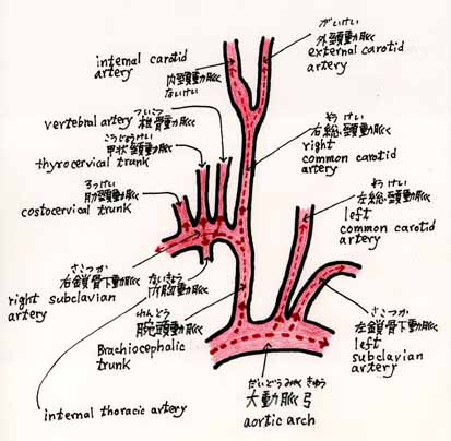 (9)長嶋茂雄氏の梗塞部位:中大脳動脈 ●長嶋茂雄氏(読売巨人軍終身名誉監督)が、体調不良を起こしたのは、3月4日(木)の早朝のことだったようです。 ●東京女子医科大学病院に緊急入院となり、内山真一郎教授(神経内科)が3月5日(金)に行った記者会見(午前11時58分・弥生記念講堂)によると、長嶋茂雄氏は、心房細動(不整脈の一種)(Atrial Fibrillation)を起して心臓内に血栓(小さい血の塊)を生じ、それが左の大脳に飛んで、脳梗塞(心原性脳塞栓症)(しんげんせい・のう・そくせんしょう)(注記)を発症したとのことです。(http://www.asahi.com/special/nagashima/TKY200403050184.html) ●1週間後の3月11日(木)に行った記者会見で、梗塞部位は中大脳動脈の根元であると発表されました。 ●左心房にできた血栓が大動脈弓から左総頸動脈に入って、内頸動脈を流れて上がり、内頸動脈の終枝(最大枝)である中大脳動脈(middle cerebral artery)を詰まらせたものと思われます。 ●内山教授の発表によると「脳の深いところで運動神経繊維が密集している場所にも、脳梗塞がおよんでいる」ようです。 ●中大脳動脈は、前交通動脈、左・右前大脳動脈、左・右内頸動脈、左・右後交通動脈、左・右後大脳動脈とともに、ウイリス動脈輪(Circle of Willis)[大脳動脈輪]を形成します。 (http://www.nlm.nih.gov/cgi/mesh/2004/MB_cgi?term=Circle+of+Willis&field=entry) ● MeSH用語では、中大脳動脈の用語は、大脳動脈の用語の下位にあります。 Cerebral Arteries(大脳動脈) (http://www.nlm.nih.gov/cgi/mesh/2004/MB_cgi?term=Cerebral+Arteries&field=entry) Scope Note: The arteries supplying the cerebral cortex. 大脳動脈は大脳皮質(だいのうひしつ)に血液を供給する動脈をいう。 Middle Cerebral Artery(中大脳動脈) (http://www.nlm.nih.gov/cgi/mesh/2004/MB_cgi?mode=&term=Middle+Cerebral+Artery&field=entry) (http://www.ncbi.nlm.nih.gov:80/entrez/query.fcgi?cmd=Retrieve&db=mesh&list_uids=68020768&dopt=Full) Scope Note: The largest and most complex of the cerebral arteries. Branches of the middle cerebral artery supply the insular region, motor and premotor areas, and large regions of the association cortex. 中大脳動脈は、大脳動脈の最大枝で多数の枝を出す複雑な動脈である。中大脳動脈の枝は、島領域(insular region)、運動野および前運動皮質野、そして連合野の大部分の領域に分布している。 (注記)心原性(しんげんせい)(cardiogenic):心臓が原因となるという意味で、脳梗塞のうち心原性のものは、全体の2〜3割で、心房細動によるものは、6〜7割を占めるといわれています。 (10)大動脈弓の枝[3]鎖骨下動脈を探る:(1)椎骨動脈(ついこつどうみゃく) ● 右側の鎖骨下動脈(さこつかどうみゃく)(Subclavian Artery)は腕頭動脈(わんとうどうみゃく)(Brachiocephalic Trunk)から、左側の鎖骨下動脈は直接、大動脈弓(だいどうみゃくきゅう)から起こります。 ● 鎖骨下動脈は、頸部、胸部および頭部の一部に血液を供給し、腋窩動脈(えきかどうみゃく)(Axillary Artery)、上腕動脈(じょうわんどうみゃく)(Brachial Artery)につながり、肘窩(ちゅうか)(上腕二頭筋腱膜の下)で橈骨動脈(とうこつどうみゃく)(Radial Artery)と尺骨動脈(しゃくこつどうみゃく)(Ulnar Artery)との2終枝に分かれます。 ● 鎖骨下動脈(Subclavian Artery)は、MeSH用語では、次ぎのように定義されています。 Subclavian Artery(鎖骨下動脈) (http://www.nlm.nih.gov/cgi/mesh/2004/MB_cgi?term=Subclavian+Artery&field=entry) Scope Note: Artery arising from the brachiocephalic trunk on the right side and from the arch of the aorta on the left side. It distributes to the neck, thoracic wall, spinal cord, brain, meninges, and upper limb. 右鎖骨下動脈は、腕頭動脈から起こり、左鎖骨下動脈は、大動脈弓から起こる。頸部、胸壁、脊髄、脳、髄膜および上肢に血液を供給する。 ● 鎖骨下動脈は、その経過中に椎骨動脈(ついこつどうみゃく)(Vertebral Artery)、内胸動脈(ないきょうどうみゃく)(internal thoracic artery)、甲状頸動脈(こうじょうけいどうみゃく)(thyrocervical trunk)、肋頸動脈(ろっけいどうみゃく)(costocervical trunk)を出します。 ● MeSHでは、このうち椎骨動脈(Vertebral Artery)だけをMeSH Headingとしており、内胸動脈(Internal Thoracic Artery)は乳腺動脈(Mammary Arteries)のエントリー・タームとなっています。(http://www.ncbi.nlm.nih.gov:80/entrez/query.fcgi?cmd=Retrieve&db=mesh&list_uids=68008323&dopt=Full) Vertebral Artery(椎骨動脈) (http://www.nlm.nih.gov/cgi/mesh/2004/MB_cgi?term=Vertebral+Artery&field=entry) Scope Note: The first branch of the subclavian artery with distribution to muscles of the neck, vertebrae, spinal cord, cerebellum and interior of the cerebrum. 椎骨動脈は、鎖骨下動脈の第1枝(最大枝)で、頸の筋肉、椎骨、脊髄、小脳および大脳の下面に分布する動脈である。 ● 頭蓋腔内(延髄と橋の移行部)で、左右両側からくる椎骨動脈は接近して1本に合流し、脳底動脈(のうていどうみゃく)(Basilar Artery)となります。 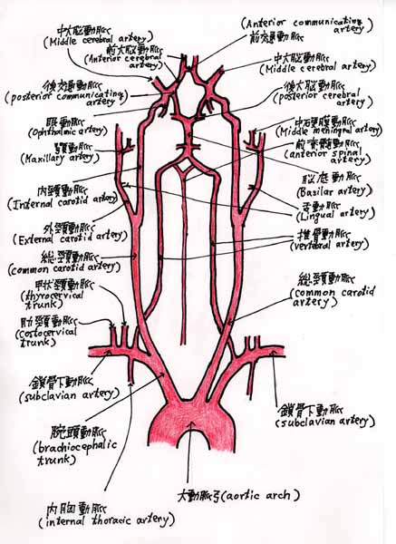 (11)大動脈弓の枝[3]鎖骨下動脈を探る:(2)脳底動脈(のうていどうみゃく) ○脳に分布する動脈には、内頸動脈の枝(前大脳動脈、中大脳動脈、前脈絡叢動脈、後交通動脈)と椎骨動脈の枝(後下小脳動脈)と椎骨動脈につづく脳底動脈の枝(前下小脳動脈、迷路動脈、橋枝、上小脳動脈、後大脳動脈)がある。(『日本人体解剖学』南山堂) 脳に分布する動脈 1. 内頸動脈の枝(大脳動脈) 前大脳動脈(anterior cerebral artery) 中大脳動脈(middle cerebral artery)(長嶋茂雄氏の梗塞部位) 前脈絡叢動脈(anterior choroid artery) 後交通動脈(posterior communicating artery) 2. 椎骨動脈の枝 後下小脳動脈(posterior inferior cerebellar artery) 3. 脳底動脈の枝 前下小脳動脈(anterior inferior cerebellar artery) 迷路動脈(labyrinthine artery) 橋枝(pontine branches) 上小脳動脈(superior cerebellar artery) 後大脳動脈(posteriior cerebral artery) ●MeSHでは、Basilar Arteryを次のように定義しています。 Basilar Artery(脳底動脈) (http://www.nlm.nih.gov/cgi/mesh/2004/MB_cgi?term=Basilar+Artery&field=entry) (http://www.ncbi.nlm.nih.gov:80/entrez/query.fcgi?cmd=Retrieve&db=mesh&list_uids=68001488&dopt=Full) Scope Note: The artery formed by the union of the right and left vertebral arteries; it runs from the lower to the upper border of the pons, where it bifurcates into the two posterior cerebral arteries. 脳底動脈は、左右の椎骨動脈が合流してつくられる動脈である。橋(きょう)(pons)の下縁から上縁に進み、2本の後大脳動脈に分岐する。 ●脳底動脈の終枝である後大脳動脈は、後交通動脈によって内頸動脈あるいは中大脳動脈と吻合することによって、大脳動脈輪(ウィリス大脳動脈輪)(Circle of Willis)をつくることになります。 (12)大脳動脈(Cerebral Arteries)の構成 ●解剖学書によると大脳動脈(Cerebral Arteries)は、内頸動脈(CAROTID ARTERY, INTERNAL)の枝である前大脳動脈(Anterior Cerebral Artery)(ぜんだいのうどうみゃく)、中大脳動脈(Middle Cerebral Artery)(ちゅうだいのうどうみゃく)、前脈絡叢動脈(anterior choroid artery)(ぜんみゃくらくそうどうみゃく)、後交通動脈(posterior communicating artery)(こうこうつうどうみゃく)をさしますが、MeSHでは、外頸動脈の枝である側頭動脈(Temporal Arteries)と脳底にいくつかの動脈が吻合してつくられるCircle of Willis(ウィリス大脳動脈輪)も大脳動脈(Cerebral Arteries)の下位に含めています。 Cerebral Arteries [A07.231.114.228](大脳動脈) Anterior Cerebral Artery [A07.231.114.228.100](前大脳動脈) Circle of Willis [A07.231.114.228.351](ウィリス大脳動脈輪) Middle Cerebral Artery [A07.231.114.228.550](中大脳動脈) Posterior Cerebral Artery [A07.231.114.228.700](後大脳動脈) Temporal Arteries [A07.231.114.228.868](側頭動脈) 1. Anterior Cerebral Artery(前大脳動脈)(ぜんだいのうどうみゃく) (http://www.nlm.nih.gov/cgi/mesh/2004/MB_cgi?mode=&term=Anterior+Cerebral+Artery&field=entry) Scope Note: Artery formed by the bifurcation of the internal carotid artery (CAROTID ARTERY, INTERNAL). Branches of the anterior cerebral artery supply the CAUDATE NUCLEUS; INTERNAL CAPSULE; PUTAMEN; SEPTAL NUCLEI; GYRUS CINGULI; and surfaces of the FRONTAL LOBE and PARIETAL LOBE. 前大脳動脈は、内頸動脈の分枝によってつくられる動脈である。前大脳動脈の枝は、尾状核(CAUDATE NUCLEUS);内包(INTERNAL CAPSULE);被殻(PUTAMEN);中隔核(SEPTAL NUCLEI);帯状回(GYRUS CINGULI)に血液を供給し、前頭葉および頭頂葉(PARIETAL LOBE)に分布する。 2. Circle of Willis(ウィリス大脳動脈輪)(だいのうどうみゃくりん) (http://www.nlm.nih.gov/cgi/mesh/2004/MB_cgi?mode=&term=Circle+of+Willis&field=entry) Scope Note: A polygonal anastomosis at the base of the brain formed by the internal carotid (CAROTID ARTERY, INTERNAL), proximal parts of the anterior, middle, and posterior cerebral arteries (ANTERIOR CEREBRAL ARTERY, MIDDLE CEREBRAL ARTERY, POSTERIOR CEREBRAL ARTERY), the anterior communicating artery and the posterior communicating arteries. ウィリス大脳動脈輪は、内頸動脈(CAROTID ARTERY, INTERNAL)、前大脳動脈、中大脳動脈、後大脳動脈の近位部分、前交通動脈および後交通動脈によって脳底つくられる多角形[六角形]吻合である。 3. Middle Cerebral Artery(中大脳動脈)(ちゅうだいのうどうみゃく) (http://www.nlm.nih.gov/cgi/mesh/2004/MB_cgi?mode=&term=Middle+Cerebral+Artery&field=entry) Scope Note: The largest and most complex of the cerebral arteries. Branches of the middle cerebral artery supply the insular region, motor and premotor areas, and large regions of the association cortex. 中大脳動脈は、大脳動脈の最大枝で多数の枝を出す複雑な動脈である。中大脳動脈の枝は、島領域(insular region)、運動野および前運動皮質野、そして連合野の大部分の領域に分布している。 4. Posterior Cerebral Artery(後大脳動脈)(こうだいのうどうみゃく) (http://www.nlm.nih.gov/cgi/mesh/2004/MB_cgi?mode=&term=Posterior+Cerebral+Artery&field=entry) Scope Note: Artery formed by the bifurcation of the BASILAR ARTERY. Branches of the posterior cerebral artery supply portions of the OCCIPITAL LOBE; PARIETAL LOBE; inferior temporal gyrus, brainstem, and CHOROID PLEXUS. 後大脳動脈は、脳底動脈(BASILAR ARTERY)の分岐によってつくられる。後大脳動脈の枝は、後頭葉(OCCIPITAL LOBE);頭頂葉(PARIETAL LOBE);下側頭回(inferior temporal gyrus);脳幹(brainstem)および脈絡叢(CHOROID PLEXUS)に血液を供給する。 5. Temporal Arteries(側頭動脈)(そくとうどうみゃく) (http://www.nlm.nih.gov/cgi/mesh/2004/MB_cgi?mode=&term=Temporal+Arteries&field=entry) Scope Note: Arteries arising from the external carotid or the maxillary artery and distributing to the temporal region. 側頭動脈は、外頸動脈あるいは顎動脈から起こる動脈である。側頭部に分布する。 ●外頸動脈から起こる側頭動脈は、浅側頭動脈(superficial temporal artery)(外頸動脈の終枝)で、主として側頭部の皮下に広がっています。 [Coffee break] Circle of Willis(ウィリス大脳動脈輪)を構成する動脈を調べておきましょう。 (13)鎖骨下動脈につづく動脈(1)腋窩動脈(Axillary Artery)と上腕動脈(Brachial Artery) ● 腋窩動脈(Axillary Artery)(えきかどうみゃく)は、鎖骨下動脈につづく動脈で、鎖骨の下縁から大胸筋下縁までの間をいいます。 ●上腕動脈(Brachial Artery)は、腋窩動脈につづく動脈で、大胸筋の下縁から肘窩(ちょうか)までをいいます。 Axillary Artery(腋窩動脈)(えきかどうみゃく) (http://www.nlm.nih.gov/cgi/mesh/2004/MB_cgi?mode=&term=Axillary+Artery&field=entry) (http://www.ncbi.nlm.nih.gov:80/entrez/query.fcgi?cmd=Retrieve&db=mesh&list_uids=68001366&dopt=Full) Scope Note: The continuation of the subclavian artery; it distributes over the upper limb, axilla, chest and shoulder. 腋窩動脈は、鎖骨下動脈につづく動脈で、上肢、腋窩、胸および肩に分布する。 Brachial Artery(上腕動脈)(じょうわんどうみゃく) (http://www.nlm.nih.gov/cgi/mesh/2004/MB_cgi?mode=&term=Brachial+Artery&field=entry) (http://www.ncbi.nlm.nih.gov:80/entrez/query.fcgi?cmd=Retrieve&db=mesh&list_uids=68001916&dopt=Full) Scope Note: The continuation of the axillary artery; it branches into the radial and ulnar arteries. 上腕動脈は、腋窩動脈につづく動脈で、橈骨動脈と尺骨動脈に分枝する。 (14)鎖骨下動脈につづく動脈(2)橈骨動脈(Radial Artery)と尺骨動脈(Ulnar Artery) ● 上腕動脈は、肘窩(ちょうか)で橈骨動脈(Radial Artery)と尺骨動脈(Ulnar Artery)とに二分します。 ●橈骨動脈は、橈骨下端の前面で皮下を走り、ここで脈がとられます。 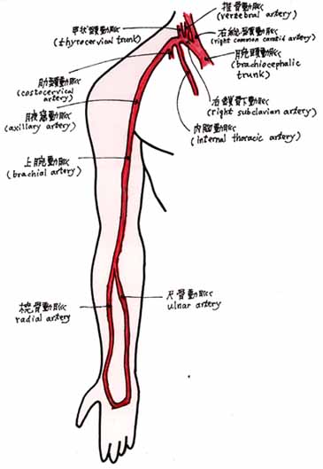 Radial Artery(橈骨動脈)(とうこつどうみゃく) (http://www.nlm.nih.gov/cgi/mesh/2004/MB_cgi?mode=&term=Radial+Artery&field=entry) (http://www.ncbi.nlm.nih.gov:80/entrez/query.fcgi?cmd=Retrieve&db=mesh&list_uids=68017534&dopt=Full) Scope Note: The direct continuation of the brachial trunk, originating at the bifurcation of the brachial artery opposite the neck of the radius. Its branches may be divided into three groups corresponding to the three regions in which the vessel is situated, the forearm, wrist, and hand. 橈骨動脈は、上腕動脈から直接つづく動脈である。橈骨頭の向かい側の上腕動脈の分岐にはじまる。橈骨動脈の枝は3群に分かれ、3つの部分(前腕、腕関節、手)に分布している。 Ulnar Artery(尺骨動脈)(しゃくこつどうみゃく) (http://www.nlm.nih.gov/cgi/mesh/2004/MB_cgi?mode=&term=Ulnar+Artery&field=entry) (http://www.ncbi.nlm.nih.gov:80/entrez/query.fcgi?cmd=Retrieve&db=mesh&list_uids=68017535&dopt=Full) Scope Note: The larger of the two terminal branches of the brachial artery, beginning about one centimeter distal to the bend of the elbow. Like the RADIAL ARTERY, its branches may be divided into three groups corresponding to their locations in the forearm, wrist, and hand. 尺骨動脈は、上腕動脈の2本の終枝のうちより太い動脈である。肘の下(遠位)の1cmほどのところにはじまる。橈骨動脈(RADIAL ARTERY)と同じように、3つの部分(前腕、腕関節、手)に分布している。 (15)大動脈の構成 ● 体循環の本幹である大動脈(Aorta)は、その部位によって、上行大動脈(Aorta, Ascending)、大動脈弓(Aorta, Thoracic)、胸大動脈(Aorta, Thoracic)、腹大動脈(Aorta, Abdominal)、正中仙骨動脈(median sacral artery)に区別されます。 左心室 ↓ 上行大動脈 → ヴァルサルヴァ洞(Sinus of Valsalva) → 冠状動脈 ↓ 大動脈弓 ↓ 胸大動脈(Aorta, Thoracic) ↓ ↓ (横隔膜)(大動脈裂孔) ↓ ↓ 腹大動脈(Aorta, Abdominal) ↓ 正中仙骨動脈 ●胸大動脈(Aorta, Thoracic)以下を下行大動脈といいます。 ●胸大動脈(Aorta, Thoracic)と腹大動脈(Aorta, Abdominal)の境が横隔膜です。 ●上行大動脈(Aorta, Ascending)は、大動脈(Aorta)のエントリー・タームです。 ●大動脈弓は、上行大動脈と下行大動脈の移行部となる彎曲部で、腕頭動脈、左総頸動脈、左鎖骨下動脈を出す大動脈の重要な部位です。 Aorta [A07.231.114.056](大動脈) Aorta, Abdominal [A07.231.114.056.205](腹大動脈) Aorta, Thoracic [A07.231.114.056.372](胸大動脈) Sinus of Valsalva [A07.231.114.056.847](ヴァルサルヴァ洞) Aorta(大動脈) (http://www.nlm.nih.gov/cgi/mesh/2004/MB_cgi?mode=&term=Aorta&field=entry) (http://www.ncbi.nlm.nih.gov:80/entrez/query.fcgi?cmd=Retrieve&db=mesh&list_uids=68001011&dopt=Full) Scope Note: The main trunk of the systemic arteries. 大動脈は、体循環(動脈系)の本幹である。 ●大動脈の起始部からは2本の冠状動脈から起こり、左右に向かい心臓全体に分布しています。 Aorta, Abdominal(腹大動脈) (http://www.nlm.nih.gov/cgi/mesh/2004/MB_cgi?mode=&term=Aorta,+Abdominal&field=entry) (http://www.ncbi.nlm.nih.gov:80/entrez/query.fcgi?cmd=Retrieve&db=mesh&list_uids=68001012&dopt=Full) Scope Note: The aorta from the diaphragm to the bifurcation into the right and left common iliac arteries. 腹大動脈は、横隔膜から左右の総腸骨動脈に分岐するまでの動脈をいう。 Aorta, Thoracic(胸大動脈) (http://www.nlm.nih.gov/cgi/mesh/2004/MB_cgi?mode=&term=Aorta,+Thoracic&field=entry) (http://www.ncbi.nlm.nih.gov:80/entrez/query.fcgi?cmd=Retrieve&db=mesh&list_uids=68001013&dopt=Full) Scope Note: The portion of the descending aorta proceeding from the arch of the aorta and extending to the diaphragm. 胸大動脈は、大動脈弓のつづきとして始まり、横隔膜までのびる下行大動脈の一部である。 ●大動脈弓(Aortic Arch)と下行大動脈(Aorta, Descending)は、胸大動脈(Aorta, Thoracic)のエントリー・タームとなっています。 Sinus of Valsalva(ヴァルサルヴァ洞) (http://www.nlm.nih.gov/cgi/mesh/2004/MB_cgi?mode=&term=Sinus+of+Valsalva&field=entry) (http://www.ncbi.nlm.nih.gov:80/entrez/query.fcgi?cmd=Retrieve&db=mesh&list_uids=68012850&dopt=Full) Scope Note: The dilatation of the aortic wall behind each of the cusps of the aortic valve. ヴァルサルヴァ洞は、大動脈弁のそれぞれの弁尖[3個の半月弁の弁膜]の大動脈壁のひろがりをいう。 ●ヴァルサルヴァ洞は、大動脈洞(aortic sinus)ともよばれ、ポッケット状にひろがっています。 (16)腹腔動脈 ○腹大動脈(abdominal aorta)上端において膵臓の上縁、すなわち第12胸椎の高さで起こり、長さ1〜2cmで、ただちに左胃動脈(left gastric artery)、総肝動脈(common hepatic artery)および脾動脈(splenic artery)の3枝に分れる。(『日本人体解剖学』) Celiac Artery(腹腔動脈) (http://www.nlm.nih.gov/cgi/mesh/2004/MB_cgi?mode=&term=Celiac+Artery&field=entry) (http://www.ncbi.nlm.nih.gov/entrez/query.fcgi?cmd=Retrieve&db=mesh&list_uids=68002445&dopt=Full) Scope Note: The arterial trunk that arises from the abdominal aorta and after a short course divides into the left gastric, common hepatic and splenic arteries 腹腔動脈は、腹大動脈から起こる動脈で、短い経過ののち、左胃動脈、総肝動脈および脾動脈に分れる。 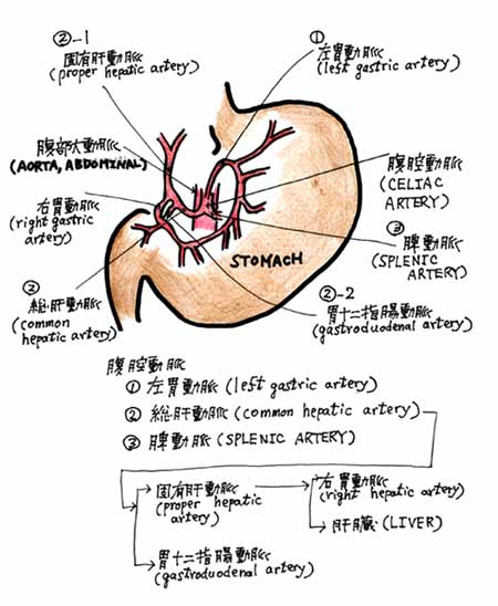 (17)脾動脈 ○「腎の後ろで膵臓の上縁に沿ってうねりながら水平に左方の進み、脾門から脾実質中に入る。」(『日本人体解剖学』南山堂) Splenic Artery(脾動脈) (http://www.nlm.nih.gov/cgi/mesh/2004/MB_cgi?mode=&term=Splenic+Artery&field=entry) (http://www.ncbi.nlm.nih.gov/entrez/query.fcgi?cmd=Retrieve&db=mesh&list_uids=68013157&dopt=Full) Scope Note: The largest branch of the celiac trunk with distribution to the spleen, pancreas, stomach and greater omentum. 脾動脈は、腹腔動脈の最大枝であり、脾臓、膵臓、胃および大網に血液を供給する。 ●MeSH用語では、腹腔動脈のことを、Celiac Arteryといいますが、この脾動脈(Splenic Artery)のスコープ・ノートのなかでは、celiac trunkという用語を使っています。 (18)腎動脈 ○ 「腎動脈(renal artery)は、上腸間膜動脈の下方1〜2cm(第2腰椎の高さ)で両側方へ直角に走り腎臓に至る。」(『日本人体解剖学』南山堂) Renal Artery(腎動脈) (http://www.nlm.nih.gov/cgi/mesh/2004/MB_cgi?mode=&term=Renal+Artery&field=entry) (http://www.ncbi.nlm.nih.gov/entrez/query.fcgi?cmd=Retrieve&db=mesh&list_uids=68012077&dopt=Full) Scope Note: A branch of the abdominal aorta which supplies the kidneys, adrenal glands and ureters. 腎動脈は、腹大動脈の枝で、腎臓、副腎および尿管に血液を供給する。 (19)総肝動脈と固有肝動脈 ● 腹腔動脈から、直接、分かれ胃・肝臓・胆嚢に向かう動脈は、総肝動脈(common hepatic artery)といい、このうち肝臓(右・左葉)に向かう動脈を固有肝動脈(proper hepatic arytery)といいます。 ● 総肝動脈(common hepatic artery)は、固有肝動脈(proper hepatic arytery)と胃十二指腸動脈(gastroduodenal artery)に分枝します。 ● MeSH用語では、総肝動脈(common hepatic artery)と固有肝動脈(proper hepatic arytery)を、肝動脈(Hepatic Artery)としています。 Hepatic Artery(肝動脈) (http://www.nlm.nih.gov/cgi/mesh/2004/MB_cgi?mode=&term=Hepatic+Artery&field=entry) (http://www.ncbi.nlm.nih.gov/entrez/query.fcgi?cmd=Retrieve&db=mesh&list_uids=68006499&dopt=Full) Scope Note: A branch of the celiac artery that distributes to the stomach, pancreas, duodenum, liver, gallbladder, and greater omentum. 肝動脈は、腹腔動脈の枝で、胃、膵臓、十二指腸、肝臓、胆嚢、および大網に血液を供給する動脈をいう。 (20)腸間膜動脈:上腸間膜動脈と下腸間膜動脈[1] ● 腹大動脈(Aorta, Abdominal)の臓側枝として出る腸間膜動脈(Mesenteric Arteries)は、上・下に分かれます。 ● 腹大動脈(Aorta, Abdominal)から出る臓側枝を、まとめておくと、以下のようになります。 1.腹腔動脈(celiac artery) 2.上腸間膜動脈(superior mesenteric artery) 3.下腸間膜動脈(inferior mesenteric artery) 4.中副腎動脈(middle suparenal artery) 5.腎動脈(renal artery) 6.精巣動脈(男性)(testicular artery)、卵巣動脈(女性)(ovarian artery) ● MeSH用語では、腸間膜動脈(Mesenteric Arteries)の下位概念に上腸間膜動脈(Mesenteric Artery, Inferior)と下腸間膜動脈(Mesenteric Artery, Superior)の用語があります。 Mesenteric Arteries [A07.231.114.565] Mesenteric Artery, Inferior [A07.231.114.565.510] Mesenteric Artery, Superior [A07.231.114.565.755] Mesenteric Arteries(腸間膜動脈) (http://www.nlm.nih.gov/cgi/mesh/2004/MB_cgi?mode=&term=Mesenteric+Arteries&field=entry) (http://www.ncbi.nlm.nih.gov/entrez/query.fcgi?cmd=Retrieve&db=mesh&list_uids=68008638&dopt=Full) Scope Note: Arteries which arise from the abdominal aorta and distribute to most of the intestines. 腸間膜動脈は、腹大動脈から起こる動脈で、大部分の腸に分布する。 21)腸間膜動脈:上腸間膜動脈と下腸間膜動脈[2] ○上腸間膜動脈:「腹腔動脈のすぐ下で大動脈から分岐する1本の太い動脈で、膵臓の後ろを通って十二指腸下部の前面に現れる。ここで腸間膜のなかにはいって著しく分岐し、主として小腸と大腸の頭側半との分布する。」(『人体解剖学』藤田恒太郎著 南江堂) Mesenteric Artery, Superior(上腸間膜動脈) (http://www.nlm.nih.gov/cgi/mesh/2005/MB_cgi?mode=&term=Mesenteric+Artery,+Superior&field=entry) Scope Note: A large vessel supplying the whole length of the small intestine except the superior part of the duodenum. It also supplies the cecum and the ascending part of the colon and about half the transverse part of the colon. It arises from the anterior surface of the aorta below the celiac artery at the level of the first lumbar vertebra. 上腸間膜動脈は、十二指腸の上部をのぞいた小腸のほとんどの部分に分布する大きな血管である。また、盲腸および上行結腸、横行結腸の約半分に血液を供給する。腹腔動脈の下で、大動脈の前面から起こり、それは、第一腰椎の高さである。 ○下腸間膜動脈:腹大動脈の下部から起こり、下行結腸・S状結腸・直腸に分布する動脈で、上腸間膜動脈より小さい。(『人体解剖学』藤田恒太郎著 南江堂) Mesenteric Artery, Inferior(下腸間膜動脈) (http://www.nlm.nih.gov/cgi/mesh/2005/MB_cgi?mode=&term=Mesenteric+Artery,+Inferior&field=entry) Scope Note: The artery supplying nearly all the left half of the transverse colon, the whole of the descending colon, the sigmoid colon, and the greater part of the rectum. It is smaller than the superior mesenteric artery (MESENTERIC ARTERY, SUPERIOR) and arises from the aorta above its bifurcation into the common iliac arteries. 下腸間膜動脈は、横行結腸の左半分のほとんど、下行結腸のすべて、S状結腸および直腸の大部分に血液を供給する動脈である。上腸間膜動脈(MESENTERIC ARTERY, SUPERIOR)より小さく、総腸骨動脈に分岐する前(上)の大動脈から起こる。 22)腸骨動脈 ●腹大動脈は、脊柱の前面を下って第4腰椎の高さで左右に総腸骨動脈を出します。 ●総腸骨動脈は、仙腸関節の高さで内腸骨動脈と外腸骨動脈とに分かれます。 →左総腸骨動脈――→左外腸骨動脈――→ | | | | 腹大動脈―――→| |→左内腸骨動脈――→ | | |→右内腸骨動脈――→ | | | | →右総腸骨動脈――→右外腸骨動脈――→ Iliac Artery(腸骨動脈) (http://www.nlm.nih.gov/cgi/mesh/2005/MB_cgi?mode=&term=Iliac+Artery&field=entry) Scope Note : Either of two large arteries originating from the abdominal aorta; they supply blood to the pelvis, abdominal wall and legs 腸骨動脈は、腹大動脈から起こる左右の2つの太い動脈ではじまり、骨盤内臓、腹壁および脚に血液を供給する。 ●総腸骨動脈は重要な解剖学用語で『解剖生理及衛生』(明治39年 南江堂書店)にも載っています。この時、外腸骨動脈の用語はありましたが、内腸骨動脈の用語はなく、下腹動脈の用語が使われていたようです。 23)大腿動脈(Femoral Artery)と膝窩動脈(Popliteal Artery) ●大腿動脈は、外腸骨動脈(がいちょうこつどうみゃく)(external iliac artery)の続きです。鼡径靱帯(そけいじんたい)(inguinal ligament)からはじまって膝関節の後面の膝窩(しつか)(popliteal fossa)で膝窩動脈となります。 Femoral Artery(大腿動脈) (http://www.nlm.nih.gov/cgi/mesh/2005/MB_cgi?mode=&term=Femoral+Artery&field=entry) Scope Note: The main artery of the thigh, a continuation of the external iliac artery. 大腿動脈は、太ももの主要な動脈であり、外腸骨動脈の続きである。 Popliteal Artery(膝窩動脈)(しつかどうみゃく) (http://www.nlm.nih.gov/cgi/mesh/2005/MB_cgi?mode=&term=Popliteal+Artery&field=entry) Scope Note: The continuation of the femoral artery coursing through the popliteal fossa; it divides into the anterior and posterior tibial arteries. 膝窩動脈は大腿動脈の続きで、膝窩(popliteal fossa)を通って、前脛骨動脈(anterior tibial artery)と後脛骨動脈(posterior tibial artery)とに分かれる。 24)脛骨動脈(けいこつどうみゃく) Tibial Arteries(脛骨動脈)(けいこつどうみゃく) (http://www.nlm.nih.gov/cgi/mesh/2005/MB_cgi?mode=&term=Tibial+Arteries&field=entry) Scope Note: The anterior and posterior arteries created at the bifurcation of the popliteal artery. The anterior tibial artery begins at the lower border of the popliteus muscle and lies along the tibia at the distal part of the leg to surface superficially anterior to the ankle joint. Its branches are distributed throughout the leg, ankle, and foot. The posterior tibial artery begins at the lower border of the popliteus muscle, lies behind the tibia in the lower part of its course, and is found situated between the medial malleolus and the medial process of the calcaneal tuberosity. Its branches are distributed throughout the leg and foot. 脛骨動脈(Tibial Arteries)は、膝窩動脈(popliteal artery)からの分枝であり、前脛骨動脈と後脛骨動脈とに分かれる。 前脛骨動脈(anterior tibial artery)は膝窩筋の下部にはじまり、脛骨に沿って、脚の遠位の部分、皮相表面、足首の関節にまで分布ている。 後脛骨動脈(posterior tibial artery)は膝窩筋の下部にはじまり、その経過中の下部の部分で脛骨の後を下って、内果(うちくるぶし)(medial malleolus)と踵骨隆起内側突起との間にみられ、下腿と足の全体に枝を出し、分布している。 (参考) ●下腿とは、膝関節と足首の間をいい、その骨格は、脛骨(けいこつ)・腓骨(ひこつ)および膝蓋骨(しつがいこつ)からなります。 ●内果の果というのは、盛り上がっている突起をいい、後頭果というような使い方をします。 25)臍動脈(さいどうみゃく) ●臍動脈は、胎生期の血液循環(fetal circulation)に関係する動脈で、胎児から炭酸ガスや老廃物を胎盤に運ぶ動脈で静脈血で満たされています。 ○「出生後は閉鎖して臍動脈索となるが、初部のみが閉鎖せず臍動脈として残り、膀胱の上部および中部に分布する上膀胱動脈を出す。」(『日本人体解剖学』下巻 南山堂) ●MeSH用語としてのUmbilical Arteriesのスコープ・ノートでは、胎児の臍動脈について説明しています。 Umbilical Arteries(臍動脈)(A07.231.114.929)(A16.254.789.641) (http://www.nlm.nih.gov/cgi/mesh/2005/MB_cgi?mode=&term=Umbilical+Arteries&field=entry) Scope Note: Either of a pair of arteries originating from the internal iliac artery and passing through the umbilical cord to carry blood from the fetus to the placenta. 臍動脈は、左右の内腸骨動脈(internal iliac artery)から起こる一対の動脈で、臍帯(さいたい)(umbilical cord)を通って、胎児から胎盤に血液を運ぶ動脈である。 26)腹壁動脈(ふくへきどうみゃく) ●動脈の旅も、そろそろ終わりに近づいてきました。このシソ−ラス研究会のホームページを、愛読してくださっている方のなかには、私が筋トレに興味を持っていることは、ご存じの方もいらっしゃるかもしれません。 ●筋トレで、腹筋を鍛えているときに、いままで腹壁動脈を意識したことはありませんでした。これからは、「腹筋の運動」をやるたびに、腹筋への神経支配や動脈の走行が気になるのかもしれません。 Epigastric Arteries(腹壁動脈) (http://www.nlm.nih.gov/cgi/mesh/2005/MB_cgi?mode=&term=Epigastric+Arteries&field=entry) Scope Note Inferior and external epigastric arteries arise from external iliac; superficial from femoral; superior from internal thoracic. They supply the abdominal muscles, diaphragm, iliac region, and groin. The inferior epigastric artery is used in coronary artery bypass grafting and myocardial revascularization. 腹壁動脈は、外腸骨動脈から起こる下腹壁動脈と外腹壁動脈、および大腿動脈から起こる浅腹膜動脈、および内胸動脈から起こる上腹壁動脈からなる。 腹壁動脈は、腹壁、横隔膜、腸骨窩および鼠径部に血液を供給する。 下腹壁動脈は、冠動脈バイパス術や心筋の再血管化ならびに再生に使用される血管である。 27)静脈弁(Venous Valves [A07.231.908.950])(New descriptor 2009)
血液〔静脈血〕を一方向にのみ流れるようにするために(逆流しないように)静脈のなかに存在する弁。通常、中程度の大きさの静脈にあり、重力に逆らって心臓に向かって血液を運ぶためにある。
(平成21年1月1日 記) |